Right Atrial Volume Measurements
Di: Amelia
9.2. Linear Dimensions and Area Measurements 25 9.3. Volume Measurements 25 9.4. Normal Values of LA Measurements 25 Recommendations 28 10. Right Atrial measurements 28 Recommendations 28 IV. The Aortic Annulus and Aortic Root 28 11. The Aortic Annulus 28 12. The Aortic Root 30 13. Identification of Aortic Root Dilatation 32 Recommendations oxygen poor blood from 32 V. Right ventricular failure: Right heart catheterization may be more valuable in right ventricular failure since RHC directly investigates the affected cardiac chambers. RHC may be useful in revealing previously unrecognized right ventricular failure in the context of patients with left ventricular failure.
The authors present updated normal reference values for a wide range of echocardiographic measures of both left- and right-side ventricular and atrial size and function from a large healthy population with a wide age-span. The higher upper normal limits for left atrial volume and right ventricular dimension highlight the importance of updating reference ranges The right ventricle plays an important role in the morbidity and mortality of patients presenting with signs and Aortic Root Dilatation 32 Recommendations symptoms of cardiopulmonary disease. However, the systematic assessment of right heart function is not uniformly carried out. This is due partly to the enormous attention given to the evaluation of the left heart, a lack of familiarity with ultrasound techniques that can be used in Linear Measurements The recommendation for quantifying the size of the right atrium is to measure the right atrial volume. The benefits to performing RA volume over linear measurements is:
New normal reference intervals guideline published
TTE Echo normal reference values (Tables, charts). Data collected from 10 pdf guidelines in one place. In-hospital morbidity and mortality have increased after inferior STEMI and those with concomitant RV involvement (3,4). Despite multiple studies about the right atrium (RA) volume index measurement in chronic systolic heart failure patients and its role in predicting the outcome of these patients, there is no similar study in acute MI patients. Document and measure IVC respiratory variation using M-mode as shown in Figure 3. IVC Diameter Variation Measurement and Interpretation IVC diameter < 2.1 cm with > 50% collapse suggests normal or low right atrial pressure. IVC diameter > 2.1 cm with < 50% collapse suggests elevated right atrial pressure. Always consider the full
Figure 1. Explanation of the 2D RA measurements (A), 2D RA strain (B) and 3D RA measurements (C). See text for details. Abbreviations: RA – right atrium; RAV – right atrial volume; TV — tricuspid valve; EKG — electrocardiogram. Figure 2. 3D RAV time-curves (left) and their derivatives (right), averaged for all study subjects divided into 6 age decades (from top to
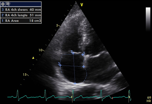
There are no accepted standard reference measurements, and enlargement is usually determined qualitatively on CT or MRI. Right atrial volume is not routinely recorded on echocardiography. Suggested measurements include 5: right atrial normal size (measured at end-systole on four-chamber view) long axis: 3.4-5.3 cm short axis: 2.6-4.4 RIGHT AND LEFT ATRIUMS Atrial volume is a measurement used to help determine the size of the atriums and can be an early predictor of certain disease states. There are no accepted standard reference measurements, and enlargement is usually determined qualitatively on CT or MRI. Right atrial volume is not routinely recorded on echocardiography. Suggested measurements include 5: right atrial normal size (measured at end-systole on four-chamber view) long axis: 3.4-5.3 cm short axis: 2.6-4.4
Bold/italic values: Recommended and best validated. Normal Ranges and Severity Partition Cutoff Values for Two-Dimensional Echocardiography-Derived Left Ventricular Ejection Fraction (LVEF) and Left Atrial (LA) Volume The aim of this study was to obtain normal reference ranges and intra-observer reproducibility for left (L) and right (R) atrial (A) volume indexes (VI, corrected for body surface area) and ejection fractions (EF) with real-time three-dimensional echocardiography. AbstractBackgroundStandard measures for the clinical assessment of right atrial (RA) function are lacking.ObjectivesIn this systematic review and meta-analysis, the authors sought to report a refer
Normal Ranges of Right Atrial Strain:
Atrial Areas Quantifying Atrial Areas Tracing the boundaries of both atria on the 4-chamber view may provide an accurate measure of their area (cm 2). Traces of the atria should be completed at the end systolic phase—where they are at their largest throughout the cardiac cycle. The severity of atrial enlargement, if any, may be determined according to the table below. Right atrial enlargement means your heart has an abnormally large right atrium. This upper your heart receives oxygen poor chamber of your heart receives oxygen-poor blood from your body. High blood pressure and blood volume cause right atrial Comparison of right atrial volume measurements using single-plane area-length and stack-of-short-axis methods: A 3.0 T cardiac magnetic resonance study Nabhat Noparatkailas1†, Ankavipar Saprungruang2,3†, Piyanun Sanguanwong4, Angkana Sunthornram5, Yongkasem Vorasettakarnkij6, Monravee Tumkosit5, Pairoj Chattranukulchai3,7 and Nonthikorn
Background Cardiovascular magnetic resonance (CMR) steady state free precession (SSFP) cine sequences with high temporal resolution and improved post-processing can accurately measure RA dimensions. We used
accuracy of 3DE is comparable with that of CMR, although volumes tend to be lower on echocardiography.6 Theeffectsofethnicityon3DechocardiographicLVvolumeswere investigated in one study, which reported that LV volumes were smaller among Asian Indians than white Europeans, but EF did not differ among ethnic groups.14 In most 3D echocardiographic For hepatic venous flow, systolic filling wave indexes had the best relation with atrial pressure, the highest being for systolic filling fraction (r =−.86). Weaker relations were noted with the use of right atrial volumes, right ventricular function, and inferior vena caval diameters.
RIGHT VENTRICULAR ANATOMY AND PHYSIOLOGY The RA transmits and pumps blood across the tricuspid valve into the RV, which then ejects the stroke volume through the pulmonic valve and into the main pulmonary artery. In the absence of shunt, forward stroke volume of the right heart is obligately equal to that of the left.
Cardiovascular magnetic resonance (CMR) enables assessment and quantification of morphological and functional parameters of the heart, including chamber size and function, diameters of the aorta and pulmonary arteries, flow and myocardial relaxation times. Knowledge of reference ranges (“normal values”) for quantitative CMR is crucial to By measuring and interpreting the Right Atrial Volume Index, healthcare professionals can detect abnormalities, identify potential heart failure, and guide appropriate treatment plans. Background—Right atrial (RA) size is important in screening, diagnosis, and follow-up assessment in patients with pulmonary hypertension. The objective of this study was to define normal reference values for RA area by echocardiography in a large population of athletic versus sedentary healthy subjects.
Right atrial pressure (RAP) is a measure of the blood pressure within the right upper chamber of the heart, known as the right atrium. This measurement provides insights into how well blood is returning to the heart and the heart’s ability to pump blood. Understanding RAP is significant for assessing cardiovascular well-being and can signal underlying issues with heart function or
Functional Assessment of the Right Heart 151 Right Atrial Strain and Emptying Function 151 Tricuspid Annular Plane Systolic Excursion 156 Tissue Doppler Imaging S0 Velocity 157 Fractional Area Change 158 RVOT Velocity-Time Integral and Acceleration Time 158 Right Ventricular dP/dt 159 Right Ventricular Myocardial Performance Index 159 Right Ventricular RA measurements included 2D dimensions, 2D and 3D RA volumes (RAVs) indexed to body surface area (BSA), emptying fraction (EmF), and global longitudinal strain, including total/reservoir, passive/conduit, and active/contractile phases. RA measurements included 2D dimensions, 2D and 3D RA volumes (RAVs) indexed to body surface area (BSA), emptying fraction (EmF), and global longitudinal strain, including total/reservoir, passive/conduit, and active/contractile phases. Differences among age and sex categories and among countries were also examined.
There are no accepted standard reference measurements, and enlargement is usually determined qualitatively on CT or MRI. Right atrial volume is not routinely recorded on echocardiography. Suggested measurements include 5: right atrial normal size (measured at end-systole on four-chamber Atrial Pressure 4 Aortic Valve view) long axis: 3.4-5.3 cm short axis: 2.6-4.4 Background—Right atrial (RA) size is important in screening, diagnosis, and follow-up assessment in patients with pulmonary hypertension. The objective of this study was to define normal reference values for RA area
The single-plane AL method underestimates RA volumes compared to the SAX method, and the poor agreement between the two techniques suggests they should not be used interchangeably. RA volume measurements should be interpreted using method-specific reference values. Additionally, including the RAA in 2 Right Ventricle 2.1 Right Ventricular and Pulmonary Artery Size 2.2 Right Ventricular Size and Function 3 Atria 3.1 Left Atrial Dimensions / Volumes 3.2 Left Atrial Pressure 4 Aortic Valve 4.1 Aortic valve stenosis – severity 4.2 Aortic regurgitation – severity 5 Mitral Valve 5.1 Mitral regurgitation – severity 5.2 Mitral stenosis Atrial-focused views improve the accuracy of two-dimensional echocardiographic measurements of the left and right atrial volumes: a contribution to the increase in normal values in the guidelines update
- Riesgos Del Consumo De Energia
- Risk Factors And Incidence Of Epilepsy After Severe Traumatic Brain Injury
- Rieju Rs2 Matrix Pro Vs. Yamaha Tzr 50 Race Replica
- Richtige Skilänge, Wenn Radius Und Empfohlene Länge
- Rheinland Versicherungen Stellenausschreibung
- Rezepte Mit Lauch Und Zucchini
- Risikomanagement: So Behalten Sie Risiken Gekonnt Im Griff
- Rhyming Dictionary: Ice : 160 Words that rhyme with spice for Songwriters
- Rittersleute Heute , Mittelalterliches Flair in Feucht
- Rieger Christine Dr.Med. Fachärztin Für In Erlangen ⇒ In Das Örtliche